Embark on a comprehensive exploration of exercise 6 classification of tissues, an essential topic in the study of biology. This in-depth analysis delves into the fundamental concepts, types, and organization of tissues, providing a solid foundation for understanding the intricate workings of living organisms.
Tissues, the fundamental building blocks of life, are classified based on their structure and function. By examining the diverse range of tissues, we gain insights into the complexity and adaptability of biological systems.
Tissue Classification Overview
Tissue classification is the systematic organization of biological tissues based on their structural and functional characteristics. It is essential for understanding the diverse cellular components of living organisms and their roles in maintaining homeostasis and performing specific functions.
Tissue is defined as a group of similar cells that perform a specific function and are organized into a structural unit. Tissues are the basic building blocks of organs, which are composed of different types of tissues working together to perform complex functions.
Criteria for Tissue Classification
The classification of tissues is based on several criteria, including:
- Cell type:Tissues are classified according to the type of cells they contain, such as epithelial cells, connective tissue cells, muscle cells, and nerve cells.
- Cell arrangement:The arrangement of cells within a tissue can vary, such as in layers, sheets, or three-dimensional networks.
- Extracellular matrix:The extracellular matrix, composed of proteins and polysaccharides, provides support and structure to tissues and can vary in composition and organization.
- Function:Tissues are classified based on their primary function, such as protection, support, movement, secretion, or absorption.
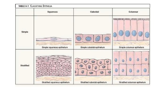
Epithelial Tissue
Epithelial tissue forms the lining of organs and cavities throughout the body, serving as a protective barrier and facilitating various functions. It is characterized by closely packed cells with minimal extracellular matrix, creating a continuous sheet-like structure.Epithelial tissues can be classified into several types based on their shape, arrangement, and function.
The primary types include:
Simple Epithelium, Exercise 6 classification of tissues
Simple epithelium consists of a single layer of cells that rests on a basement membrane. It is commonly found in areas where absorption, secretion, or filtration occurs, such as the lining of the small intestine, capillaries, and alveoli in the lungs.
Stratified Epithelium
Stratified epithelium comprises multiple layers of cells, with the basal layer attached to the basement membrane and the superficial layer facing the lumen. It provides protection in areas subjected to wear and tear, such as the skin, esophagus, and vagina.
Pseudostratified Epithelium
Pseudostratified epithelium appears to be stratified but is actually composed of a single layer of cells. The nuclei of the cells are located at different levels, giving the impression of multiple layers. It is found in areas where both secretion and protection are required, such as the lining of the trachea and bronchi.
Glandular Epithelium
Glandular epithelium consists of cells specialized for secretion. It forms glands that release various substances, such as hormones, enzymes, and mucus. Exocrine glands secrete their products into ducts that lead to the body’s surface or internal cavities, while endocrine glands secrete directly into the bloodstream.
Sensory Epithelium
Sensory epithelium contains specialized cells that detect stimuli such as light, sound, or touch. It is found in sensory organs, such as the eyes, ears, and taste buds.
Connective Tissue
Connective tissue is a type of tissue that connects, supports, and protects other tissues in the body. It is the most abundant tissue in the body, and it is found in all organs and tissues.
Connective tissue is characterized by its high content of extracellular matrix (ECM). The ECM is a complex network of proteins, polysaccharides, and water that provides structural support and protection for cells.
Types of Connective Tissue
There are three main types of connective tissue:
- Loose connective tissue
- Dense connective tissue
- Specialized connective tissue
Loose Connective Tissue
Loose connective tissue is the most common type of connective tissue. It is found in all organs and tissues, and it provides support and protection for cells.
Loose connective tissue is characterized by its high content of ECM. The ECM is a loose network of proteins, polysaccharides, and water that allows cells to move and exchange nutrients and waste products.
Dense Connective Tissue
Dense connective tissue is a type of connective tissue that is characterized by its high density of ECM. The ECM is a dense network of proteins, polysaccharides, and water that provides strong support and protection for cells.
Dense connective tissue is found in tendons, ligaments, and bones.
Specialized Connective Tissue
Specialized connective tissue is a type of connective tissue that has a specific function. Specialized connective tissue includes cartilage, bone, and blood.
Cartilage is a type of connective tissue that is found in joints and between bones. It provides support and cushioning for bones.
Bone is a type of connective tissue that is found in the skeleton. It provides support and protection for the body.
Blood is a type of connective tissue that is found in the circulatory system. It transports oxygen, nutrients, and waste products throughout the body.
Muscle Tissue
Muscle tissue is a specialized type of tissue that is responsible for movement and locomotion. It is characterized by the presence of contractile proteins, which allow it to shorten and lengthen in response to stimuli.
There are three main types of muscle tissue: skeletal muscle, smooth muscle, and cardiac muscle. Each type has a distinct structure and function.
Skeletal Muscle
Skeletal muscle is attached to bones and is responsible for voluntary movement. It is composed of long, cylindrical fibers that are multinucleated. Skeletal muscle is striated, meaning that it has a banded appearance when viewed under a microscope.
Skeletal muscle is the most abundant type of muscle tissue in the body. It is responsible for a wide range of movements, from simple actions like walking and talking to complex movements like running and jumping.
Smooth Muscle
Smooth muscle is found in the walls of hollow organs, such as the stomach, intestines, and blood vessels. It is composed of spindle-shaped fibers that are uninucleated. Smooth muscle is non-striated, meaning that it does not have a banded appearance when viewed under a microscope.
Smooth muscle is responsible for involuntary movements, such as the peristalsis of the intestines and the constriction of blood vessels. It is also responsible for maintaining the tone of hollow organs.
Cardiac Muscle
Cardiac muscle is found only in the heart. It is composed of branched fibers that are uninucleated. Cardiac muscle is striated, meaning that it has a banded appearance when viewed under a microscope.
Cardiac muscle is responsible for the rhythmic contractions of the heart. It is the only type of muscle tissue that is capable of spontaneous contraction.
Nervous Tissue
Nervous tissue is a specialized type of tissue that forms the nervous system. It is responsible for receiving, processing, and transmitting information throughout the body. Nervous tissue is composed of two main types of cells: neurons and neuroglia.Neurons are the functional units of the nervous system.
They are responsible for transmitting electrical and chemical signals throughout the body. Neurons have a cell body, dendrites, and an axon. The cell body contains the nucleus and other organelles. Dendrites are short, branched extensions of the cell body that receive signals from other neurons.
The axon is a long, thin extension of the cell body that transmits signals to other neurons or to muscles and glands.Neuroglia are cells that support and protect neurons. They make up about 90% of the cells in the nervous system.
There are several different types of neuroglia, each with a specific function. Astrocytes are star-shaped cells that provide structural support for neurons and help to maintain the blood-brain barrier. Oligodendrocytes are cells that wrap around the axons of neurons and provide insulation.
Schwann cells are cells that wrap around the axons of neurons in the peripheral nervous system. Microglia are cells that remove debris and pathogens from the nervous system.Nervous tissue is found throughout the body. It is found in the brain, spinal cord, and peripheral nerves.
The brain is the central processing unit of the nervous system. It is responsible for receiving, processing, and transmitting information from all over the body. The spinal cord is a long, thin bundle of nerves that runs from the brain down the back.
It carries signals between the brain and the rest of the body. Peripheral nerves are nerves that branch out from the spinal cord and innervate the muscles, glands, and organs of the body.
Tissue Organization
Tissue organization refers to the arrangement and structural relationships between cells and extracellular matrix within a tissue. It involves the coordination and integration of cells to perform specific functions.
There are different levels of tissue organization, each with increasing complexity and specialization:
Cellular Level
At the cellular level, individual cells exhibit specific shapes, sizes, and functions. They interact with each other through cell junctions and extracellular matrix.
Tissue Level
Tissues are groups of similar cells that perform a specific function. They can be classified into four main types: epithelial, connective, muscle, and nervous tissue.
Organ Level
Organs are composed of two or more different tissues that work together to perform a specific function. For example, the heart is composed of muscle, connective, and nervous tissue.
Organ System Level
Organ systems are groups of organs that work together to perform a specific function. For example, the digestive system consists of the stomach, intestines, and other organs that work together to digest food.
Examples of Tissue Organization in Organs
- Skin:Epidermis (epithelial), dermis (connective), hypodermis (connective)
- Heart:Myocardium (muscle), endocardium (epithelial), epicardium (connective)
- Stomach:Mucosa (epithelial), submucosa (connective), muscularis externa (muscle), serosa (epithelial)
Top FAQs: Exercise 6 Classification Of Tissues
What is the significance of tissue classification?
Tissue classification provides a systematic framework for understanding the diverse range of tissues in living organisms. It enables researchers and medical professionals to study and diagnose diseases, develop treatments, and gain insights into the fundamental principles of biology.
How are tissues classified?
Tissues are classified based on their structure and function. The four main types of tissues are epithelial, connective, muscle, and nervous tissues. Each type has distinct characteristics and performs specific roles within the body.
What is the role of epithelial tissue?
Epithelial tissue forms the lining of organs and cavities, providing protection, secretion, and absorption. It is composed of closely packed cells that form a continuous layer.